A 65 F WITH STATUS EPILEPTICUS AND K/C/O EPILEPSY SINCE 21 YEARS
This is an online e log book to discuss our patient de-identified health data shared after taking his / her / guardians signed informed consent. Here we discuss our individual patients problems through series of inputs from available global online community of experts with an aim to solve those patients clinical problem with collective current best evident based input.
This E blog also reflects my patient centered online learning portfolio and your valuable inputs on the comment box is welcome.
I have been given this case to solve in an attempt to understand the topic of " patient clinical data analysis" to develop my competency in reading and comprehending clinical data including history, clinical findings, investigations and come up with diagnosis and treatment plan.
65 years old female came to casualty with
C/C of headache since 4 hours and
Involuntary movements in right upper and lower limbs since 3 hours
Hopi:
Patient was apparently
asymptomatic 4 hours back then she developed headache in frontal region, sudden in onset, took seizure medication levipil 500 mg. Involuntary movements in right upper and lower limbs since 3 hours, 2 episode every 5 mins, each episode lasted for 2-3 mins.
Associated with uprolling of eyeballs, frothing from the mouth, involuntary defection and is unresponsive since 3 hours
No H/O fever or vomitings
PAST HISTORY:-
Known case of seizure disorder since 21 years, taking tab. Levipil 500 mg.
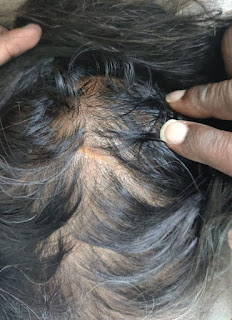
H/O head surgery for meningioma 21 years back.
Scar from the head surgery
N/k/c/o DM, HTN, Asthma, CAD, CVA, TB
Active seizure episode when she was brought to casualty:
GENERAL PHYSICAL EXAMINATION
Pt is on ventilator
pallor, icterus, cyanosis, clubbing, lymphadenopathy, edema were absent.
Bp: 120/70 mmHg
PR: 90 bpm
SpO2: 100% on FiO2 100
SYSTEMIC EXAMINATION:
CVS:
Inspection:
There are no chest wall abnormalities
The position of the trachea is central.
Apical impulse is not observed.
There are no other visible pulsations, dilated and engorged veins, surgical scars or sinuses.
Palpation:
Apex beat was localised in the 5th intercostal space 2cm lateral to the mid clavicular line
Position of trachea was central
There we no parasternal heave , thrills, tender points.
Auscultation:
S1 and S2 were heard
There were no added sounds / murmurs.
Respiratory system:
Bilateral air entry is present
Normal vesicular breath sounds are heard.
Per Abdomen:
Shape is scaphoid
Abdomen is soft and non tender with no signs of organomegaly
Bowel sounds are heard
CNS:
GCS :- E2V1M4
HIGHER MENTAL FUNCTIONS- couldn't be elicited
Memory :- couldn't be elicited
CRANIAL NERVES : couldnt be assessed
SENSORY EXAMINATION :- Could not be performed
MOTOR EXAMINATION
Normal tone in upper and lower limb
Power:- couldn't be assessed
Gait: couldnt asses the patient was ventilated
REFLEXES
Normal, brisk reflexes elicited- biceps, triceps, knee and ankle reflexes elicited
CEREBELLAR FUNCTION :- couldn't be elicited
Provisional diagnosis: STATUS EPILEPTICUS
FOCAL SEIZURES WITH SECONDARY GENERALISATION WITH TYPE 2 RESPIRATORY FAILURE SECONDARY TO POST ICTAL CONFUSION
MRI:-
EEG:-
Investigations:-
Chest X Ray
8/5/23
9/5/23
X ray pelvis AP view
Lab Investigations
Hemogram:-
8/5/23
HEMOGLOBIN- 13.9
PCV - 42.1
TLC -10 200
RBC -4.83
PLATELET COUNT - 2.44LAKH
9/5/23
HEMOGLOBIN - 13.9
PCV -37.6
TLC -9500
RBC -4.36
PLATELET COUNT -2.23LAKH
ABG:-
8/5/23 8:40 am 5:10 pm( pt was intubated)
pH -> 7.07 7.4
pCO² -> 95.3 33.6
pO² -> 114 282
HCO³‐ -> 26.4 20.4
9/5/23 7:40 am 6:40 pm
pH -> 7.43 7.48
pCO² -> 30.1 28.4
pO² -> 124 93.2
HCO³‐ -> 19.7 21.3
10/5/23
pH -> 7.46
pCO² -> 31.5
pO² -> 74.1
HCO³‐ -> 22.4
RFT 8/5/23 9/5/23 10/5/23 11/5/23
B. Urea 31 l32
Sr.Creat 0.8 0.9
Na+ 142 140 137 143
K+ 4.2 4.0 3.6 3.6
Cl- 104 104 103 104
LFT
T. Bil 1.07
D. Bil 0.22
SGOT 37
SGPT 19
Alk. Phos 179
COURSE IN THE HOSPITAL:-
A 65 YEAR OLD FEMALE PRESENTED WITH ABOVE MENTIONED COMPLAINTS AND WAS EVALUATED CUNICALLY AND WITH APPROPRIATE INVESTIGATIONS, PATIENT WAS DIAGNOSED WITH STATUS EPILEPTICUS SECONDARY TO LEFT PARIETAL
ENCEPHALOMALACIA FOCAL SEIZURES SECONDARY TO GENERALISATION
PATIENT PRESENTED TO CASUALITY WITH STATUS EPILEPTICUS AND WAS MANAGED
CONSERVATIVELY INIO REFRACTORY SEIZURES AND TYPE 2 RESPIRATORY FAILURE
EMERGENCY INTUBATION WAS DONE
MRI WAS DONE AND ENCEPHALOMALACIA IN THE LEFT PARIETAL REGION WAS NOTICED
PATIENT WAS ON MIDAZOLAM INFUSION FOR 1 AND HALF DAY AND INFUSION WAS
TAPERED AND MECHANICAL VENTILALƏN SETTINGS WERE CHANGED FROM
ACMV--->CPAP---> WAS EXTUBATED THE FOLLOWING DAY ---> NO FURTHER SEIZURE ACTIVITY WAS NOTICED PATIENT WAS ON DUALANTI EPILEPTIC INJ. LEVITARECITAM AND INJ. SODIUM VALPROATE, THEN CHANGED TO DUAL ANTIEPILEPTIC THERAPY ON TAB. LEVITARECITAM AND TAB. SODIUM VALPROATE
EEG WAS DONE WHICH SHOWED NO ABNORMALITIES AND NEUROLOGIST OPINION WAS TAKEN AND ADVICED TO CONTINUE
SAME TREATMENT
GENERAL SURGERY OPINION WAS TAKEN I/O GRADE 1 BED SORE AND ADVICED
REGULAR DRESSING WITH NEOSPORIN
PATIENT HAS RECOVERED SYMPTOMATICALLY AND DISCHARGED IN STABLE CONDITION
FINAL DIAGNOSIS:-
1)STATUS EPILEPTICUS(RESOLVED) SECONDARY TO LEFT PARIETAL ENCEPHALOMALACIA
FOCAL SEIZURES With 2⁰ GENERALIZATION
2)TYPE 2 RESPIRATORY FAILURE(RESOLVED) SECONDARY TO POST ICTAL CONFUSION
3)DENOVO HBSAG(POSITIVE) WITHSP POST EXTUBATION ON 10/5/23(9:30AM) KNOWN
CASE OF SEIZURES SINCE 16YEAR
4)HISTORY OF SURGERY FOR MENINGIOMA IN 2007
09/05/23
TREATMENT:
1. INJ. LEVIPIL 1gm IV BD
2. IV FLUIDS NS/RL @75ML/HR
3. INJ SODIUM VALPROATE 500 MG IN 100 ML NS IV INFUSION BD
4. INJ. MIDAZOLAM 8 mg/Hr IV INFUSION
5. RT FEEDS- 100 ml MILK 4th HRLY, 100 ml WATER 2nd HRLY
6. ABG 12th HLRY
7. STRICT I/O CHARTING
8. MONITOR VITALS HOURLY BP/PR/SPO2/RR
9. FREQUENT POSITION CHANGING
10/05/23
1. INJ. LEVIPIL 1gm IV BD
2. IV FLUIDS NS/RL @75ML/HR
3. INJ SODIUM VALPROATE 500 MG IN 100 ML NS IV INFUSION BD
4. INJ. MIDAZOLAM 8 mg/Hr IV INFUSION
5. RT FEEDS- 100 ml MILK 4th HRLY, 100 ml WATER 2nd HRLY
7. STRICT I/O CHARTING
8. MONITOR VITALS HOURLY BP/PR/SPO2/RR
9. FREQUENT POSITION CHANGING
11/05/23
- INJ. LEVIPIL 1gm IV BD
- IV FLUIDS NS/RL @75ML/HR
- INJ SODIUM VALPROATE 500MG IN 100 ML NS IV INFUSION BD
- NEOSPORIN DRESSINGS FOR BED SORE.
12/05/23
- INJ. LEVIPIL 1gm IV BD
- IV FLUIDS NS/RL @75ML/HR
- INJ SODIUM VALPROATE 500MG IN 100 ML NS IV INFUSION BD
- NEOSPORIN DRESSINGS FOR BED SORE.
13/05/23
- TAB SODIUM VALPROATE 200MG PO BD
- TAB LEVIPIL 500MG PO/BD
- INJ. DICLOFENAC IM SOS
- TAB PAN 40MG
- NEOSPORIN DRESSINGS FOR BED SORE.
14/05/23
- TAB SODIUM VALPROATE 200MG PO/BD
- TAB LEVIPIL 500MG PO/BD
- TAB BENFOTHIAMINE 100MG PO/ OD
- TAB PAN 40MG
- NEOSPORIN DRESSINGS FOR BED SORE.
Case Discussion:-
[08/05, 21:13] Dr. Rakesh Biswas: ABG repeated?
Sedation rate? Share the complete details of ventilation settings and current pharmacological dose and drip rate
[10/05, 21:33] Dr. Ajay: https://pubmed.ncbi.nlm.nih.gov/20581136/
[10/05, 21:36] Rakesh Biswas Sir GM HOD: So with this information what does your patient fit into?
[10/05, 21:36] Dr. Ajay: https://go.gale.com/ps/i.do?p=AONE&u=googlescholar&id=GALE|A128075131&v=2.1&it=r&sid=AONE&asid=7d1af1d7
[10/05, 21:37] Dr. Ajay: Source
[10/05, 21:40] Dr. Ajay: The patient fits mostly into true seizures sir
The patient doesn't have any intention to do pseudoseizures as per the history provided by the attenders and patient ,
There is rigidity f/b jerking,
There is involuntary defecation and micturition
[10/05, 22:50] Dr. Rakesh Biswas: Any review of literature around respiratory and metabolic acidosis in seizures similar to what our patient had?
[11/05, 08:35] Dr. Navya : https://www.ncbi.nlm.nih.gov/books/NBK441871/
Treatment of PNES may be difficult, but it is clear that anti-epileptic drugs (AEDs) are of no benefit. In addition to unnecessary costs and the potential side effects of AEDs for these patients, life-threatening side effects such as respiratory depression may occur if psychogenic nonepileptic status epilepticus is treated with large dosages of benzodiazepines.
[11/05, 09:19] Dr. Ajay: https://linkinghub.elsevier.com/retrieve/pii/S0007-0912(17)47615-X
[11/05, 09:25] Dr. Ajay : https://pubmed.ncbi.nlm.nih.gov/1191463/
[11/05, 11:57] Dr. Rakesh Biswas: Share the images of encephalomalacia from this patient's MRI as well as the previous patient with migraine and visual loss asap @Dr. Navya Ma’am PG GM
[14/05, 08:54] Dr. Rakesh Biswas: 👆MRI images of encephalomalacia ? @Dr. Deepika Ch GM PG Ma'am
[15/05, 23:59] Dr. Deepika: Age, number of lesion sites, size of encephalomalacia, and seizure frequency were independent risk factors for the prognosis of patients with REAE (OR > 1, P < 0.05). Surgical treatment was an independent protective factor associated with the prognosis of patients with REAE
variety of causes can lead to liquefaction and necrosis of brain tissue and the formation of encephalomalacia [9]. These causes include trauma, cerebrovascular disease, and intracranial infection [10]. The pathological manifestations of brain soft focus ranged from early neuronal necrosis to neuronal disappearance and then to glial cell proliferation. There are no nerve cells in the brain softening focus, which does not cause epileptic discharge. The real pathological site of epileptic discharge is the peripheral nerve tissue [11]. The traction of fibrous scar tissue in the brain can embed the remaining normal neurons cause abnormal discharge and disrupt the function of intertwined proliferative cells. It affects the electrical activity of normal neurons, resulting in seizures. A study suggested that glial cells can lead to epileptic seizures through mechanisms such as increasing the excitability of normal neurons, neuronal cluster discharge, and failure to inhibit the excitability of neurons
[15/05, 23:59] Dr. Deepika: https://www.ncbi.nlm.nih.gov/pmc/articles/PMC9273423/
[16/05, 00:00] Dr. Deepika: The predictive efficacy of encephalomalacia size on the prognosis of patients with refractory epilepsy secondary to encephalomalacia. AUC: area under the curve; CI: confidence interval.
[16/05, 00:00] Dr. Deepika: Efficacy of seizure frequency in predicting prognosis in patients with refractory epilepsy secondary to encephalomalacia. AUC: area un
[16/05, 00:02] Dr. Deepika: refractory epilepsy associated with encephalomalacia (REAE).
[16/05, 06:47] Rakesh Biswas Sir GM HOD: For the AJND article :
Can encephalomalacia be a form of neurodegeneration?
[16/05, 10:39] Dr. Deepika: Encephalomalacia also known as cerebromalacia, is the softening of brain tissue. It can be caused either by vascular insufficiency, and thus insufficient blood flow to the brain, or by degeneration. Encephalomalacia can be the formation of necrosis, or dead tissue, in a portion of the brain due to a partial complete blockage of blood flow to the area, which in turn can be caused by a natural condition or by infection or trauma (TBI). The term encephalomalacia is also used at times to refer more generally to degenerative conditions affecting the brain. If the condition affects the white matter of the brain, it is called leukoencephalomalacia. If it affects the gray matter, it is known as polioencephalomalacia.
[16/05, 10:40] Dr. Deepika Ch GM PG Ma'am: https://www.passenpowell.com/encephalomalacia-brain-injury-children-adults/
[17/05, 07:18] Dr. Rakesh Biswas: What about microscopic description of the pathology in encephalomalacia?
Also focus on it's association with vascular neurodegeneration
Also share the MR images of the 35F with CRVO and Vascular migraine and encephalomalacia
[20/05, 13:14] Dr. Deepika: CAA has now been linked with brain atrophy in regions remote from those directly affected by intracerebral hematomas, and with risk for progressive cognitive decline in the absence of new hemorrhagic strokes. Therefore, CAA is associated with features – brain atrophy and progressive cognitive decline – that are typically considered hallmarks of neurodegenerative disease. Although CAA is usually accompanied by some degree of Alzheimer's disease pathology, the profiles of cortical thinning and cognitive impairment do not fully overlap with those seen in Alzheimer's disease, suggesting that there are CAA-specific pathways of neurodegeneration. CAA-related brain ischemia may be an important mechanism that leads to brain injury, cortical disconnection, and cognitive impairment
The terms ‘neurodegeneration’ and ‘neurodegenerative’ are widely used but without a formal definition in the peer-reviewed literature. The terms are frequently used to refer to a group of neurological conditions marked by death of neurons and supporting cells in the neurovascular unit, with accompanying progressive loss of cognitive and motor functions.
Stroke and other forms of monophasic acute brain injury (e.g., because of trauma) are not typically considered to be neurodegenerative disorders. Therefore, CAA is most commonly conceptualized as a cerebrovascular disease associated with risk of lobar ICH, and not as a cause of neurodegeneration. However, accruing evidence suggests that CAA is not so easy to pigeonhole as either a cause of stroke or neurodegeneration, but instead is an important cause of both of these neurological syndromes. To make my case that CAA should be considered a neurodegenerative disease, I focus on associations between CAA and two cardinal features of neurodegenerative diseases: cognitive decline and brain atrophy
Both are a consequence of cleavage of the amyloid precursor protein to form pathogenic Aβ which aggregates into β-amyloid. In the case of AD pathology, the β-amyloid aggregates in the brain parenchyma in the form of neuritic plaques. In the case of CAA, the β-amyloid aggregates in the walls of small arteries and arterioles. Most patients with CAA also have some degree of neuritic plaques, and most patients with AD have some degree of CAA
pattern of cortical thinning in CAA (Fotiadis et al. 2016) overlaps with AD (Dickerson et al. 2009) in some regions (supramarginal gyrus, superior frontal gyrus, and inferior temporal gyrus), but other regions exhibit more thinning in CAA than AD (occipital cortex and medial frontal cortex) or in AD than CAA (precuneus, angular gyrus, and anterior temporal cortex) (Fig. 2). Overall, there are clear differences between the CAA and AD patterns of cortical thinning.
These differences in cognitive profile and regional cortical thinning suggest that neurodegeneration in CAA cannot be solely attributed to the effects of concomitant AD pathology.


Comments
Post a Comment